A biopsy is the definitive method for diagnosing mesothelioma, as it involves obtaining and analyzing tissue samples. This section explores various biopsy techniques, each chosen based on the suspected mesothelioma type and location.
Overview of Key Biopsy Techniques
Needle Biopsy
Procedure: In a needle biopsy, doctors use a thin needle to extract a small sample of tissue. This procedure is minimally invasive and is often performed under local anesthesia with imaging guidance, such as CT or ultrasound, to ensure accuracy.
Benefits: Needle biopsies are particularly useful for diagnosing pleural mesothelioma or other tumors close to the surface, as they involve minimal recovery time. However, larger tissue samples are often needed to confirm mesothelioma accurately, so needle biopsies may be used as an initial step.
👉 Learn more about supporting diagnostic tools like imaging techniques.
Thoracoscopy
Procedure: Thoracoscopy is a minimally invasive surgical procedure used to diagnose pleural mesothelioma. It involves making a small incision in the chest wall, through which a thin, camera-equipped tube (thoracoscope) is inserted to examine the pleura directly. During this procedure, doctors can obtain larger tissue samples for analysis.
Benefits: Thoracoscopy provides direct visualization of the chest cavity, allowing doctors to locate and sample suspicious areas with high accuracy. It is often chosen when pleural mesothelioma is suspected, as it provides larger tissue samples for a definitive diagnosis.
Laparoscopy
Procedure: Similar to thoracoscopy but focused on the abdomen, laparoscopy involves making a small incision to insert a camera-equipped tube into the abdominal cavity. This technique is used primarily for diagnosing peritoneal mesothelioma, allowing doctors to inspect the peritoneal lining and obtain tissue samples.
Benefits: Laparoscopy offers a minimally invasive way to access the abdominal cavity and obtain tissue samples directly from the peritoneum. It is effective for detecting mesothelioma in the abdominal lining and can help doctors understand the extent of the disease.
Mediastinoscopy
Procedure: Mediastinoscopy is used to examine lymph nodes in the chest when mesothelioma has potentially spread beyond the primary tumor. A small incision is made at the base of the neck, and a tube is inserted to reach lymph nodes, where tissue samples are taken.
Benefits: This technique is valuable for staging, as lymph node involvement typically indicates more advanced disease. It’s an effective tool for assessing whether pleural mesothelioma has spread to other areas within the chest.
Pathology and Subtype Identification
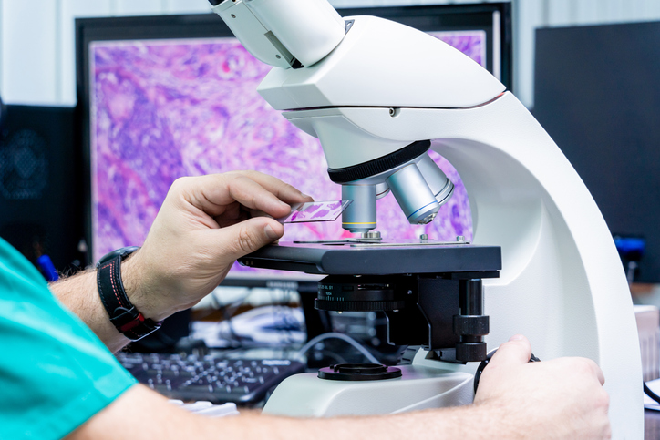
After samples are obtained, they undergo microscopic examination to confirm the presence of mesothelioma cells. Additional tests, such as immunohistochemistry, may be performed to differentiate mesothelioma from other cancers, providing clarity on the subtype (epithelioid, sarcomatoid, or biphasic) and helping to tailor the treatment plan accordingly.
👉 For a full overview of the diagnostic process, visit Diagnostic Procedures and Tests.
👉 For more about how imaging supports diagnosis, see our page on Imaging Techniques.